
中府 (Zhōngfǔ) — Central Treasury
Lung channel · point 1
Traditionally used for
- Cough & breathing
- Pain & joints
Cautions & contraindications
- No deep needling
Location
On the anterior thoracic wall, level with the first intercostal space, lateral to the infraclavicular fossa, 6 cun lateral to the anterior midline; 1 cun inferior to LU2.
How to locate: Find the depression below the lateral end of the clavicle (LU2), then drop one cun to the first intercostal space. It lies on the same transverse line along the first intercostal space as ST14, KI26 and CV20, not on the ribs themselves.
Regional anatomy: Superficial to the pectoralis major and minor; the lung apex lies deep to this region, which is why the needle is angled away from the chest wall.
Distances are in cun, a proportional unit — how to measure.
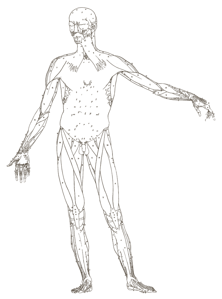
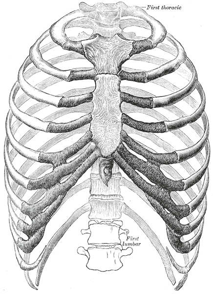 Needling hazard — see Needling safety
3D view
3D view not available for this point: its position can't be carried onto the 3D model reliably (it failed a placement check). The 2D maps above show it.
- The lung is not drawn: the anatomical model used here does not include it as a surface. This is a limit of the model, not of the anatomy. The pneumothorax risk above still applies.
Needling
Cited needling
- ⚠ Pneumothorax
-
Transverse-oblique insertion — medially along the intercostal spaceDepth 0.5–1 cunskindeeper
Depths describe average adult habitus in the cited population, never a single safe number for the patient in front of you. Palpate and adjust.
Sources (2)
- World Health Organization. Guidelines on basic training and safety in acupuncture. Geneva: WHO; 1999. WHO/EDM/TRM/99.1 — Section II §5.2 (Lung and pleura), p. 25 Supports: pneumothorax (names this region)
- Deadman P, Al-Khafaji M, Baker K. A Manual of Acupuncture. Hove: Journal of Chinese Medicine Publications; 1998 — p. 76 Supports: techniques (names this point)
Site notes · not cited
- No deep needling
Functions
- Disseminates and descends Lung qi
- Transforms phlegm and clears heat from the chest and upper burner
- Regulates the water passages
- Harmonises the Lung and Spleen
- Moves qi locally and in the sinew channel
Indications
Clinical Notes
Similar Points
Ranked by shared patterns and conditions
Sources
- World Health Organization. WHO Standard Acupuncture Point Locations in the Western Pacific Region. Manila: WHO Regional Office for the Western Pacific, 2008. (Location.)
- Focks C, ed. Atlas of Acupuncture. Edinburgh: Churchill Livingstone Elsevier, 2008. (Locating method and actions.)
📝 Notes
Public notes from the community and your own private notes on LU1 Zhongfu.
No notes yet.