
Traditionally used for
- Nerves & recovery
- Pain & joints
Location
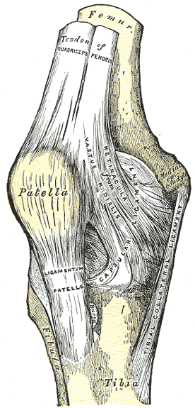
On the knee, in the depression lateral to the patellar ligament, with the knee flexed.
How to locate: Flex the knee to 90°; the point is the lateral of the two hollows ('the calf's nostrils') below the kneecap.
Distances are in cun, a proportional unit — how to measure.
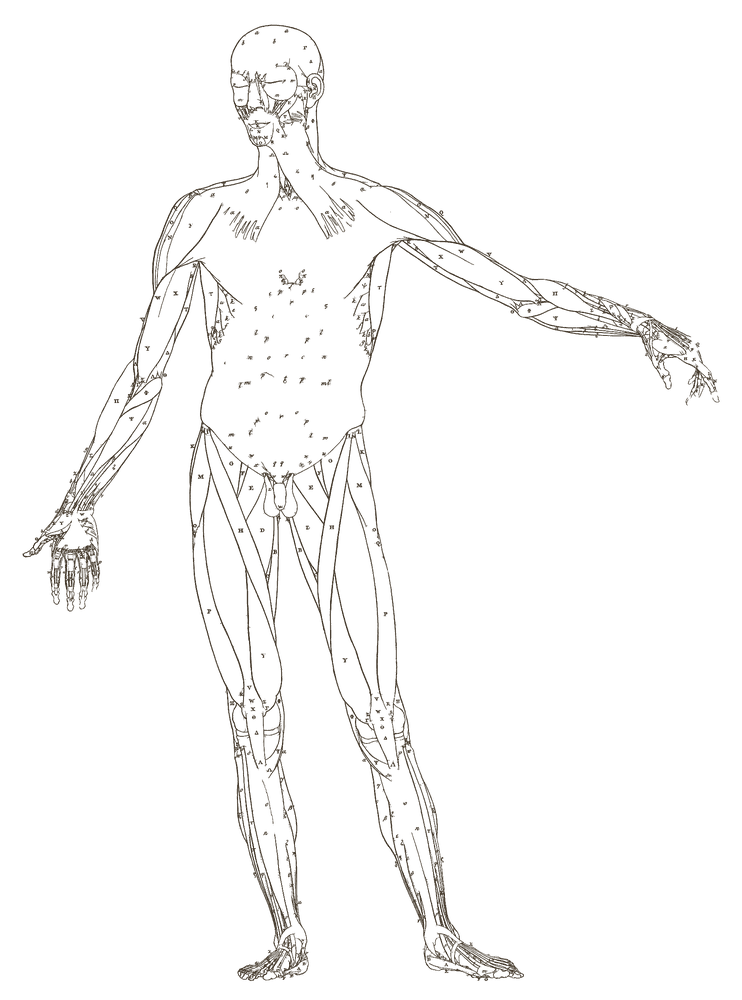
3D view
The 3D view is not available on this device. The 2D maps above show the same point.
Drag, or focus the model and use the arrow keys, to rotate; + and − to zoom; 0 to reset; Esc to close.
Needle diagram for the marked point — a schematic section through the skin, not the model.
3D anatomy: BodyParts3D, © The Database Center for Life Science licensed under CC Attribution 4.0 International (licence). Subset, simplified and recoloured by Verscienta — changes and licence details.
Approximate placement, carried over from our 2D body map onto a single adult male model (BodyParts3D) and checked against the point's location text. Shown on one side of the body. Locate the point on your patient by the location text and palpation, not by this model.
- Cun on this part of the body
- From the superior border of the pubic symphysis to the base of the patella: 18 B-cun (WHO 2008, General Guidelines, Proportional Bone (Skeletal) Measurements, p. 13).
- Needle angle and depth
-
- Perpendicular (90° to the skin), 1–2 cun, directed towards Weizhong BL40. Directed towards BL40. Deadman P, Al-Khafaji M, Baker K, p. 157
- Oblique (45° to the skin), 1–2 cun, in a medial and superior direction, behind the patella. The direction is given in the source's words only. Deadman P, Al-Khafaji M, Baker K, p. 157
- Through needling, behind the patellar ligament to medial xiyan (mn-le-16) — not drawn: no cited depth in cun.
3D anatomy: BodyParts3D, © The Database Center for Life Science licensed under CC Attribution 4.0 International (licence). Subset, simplified and recoloured by Verscienta — changes and licence details.
Needling
Cited needling
Techniques — the source gives these as alternatives:
-
Perpendicular insertion — directed towards Weizhong BL40Depth 1–2 cunskindeeper
With the knee flexed and supported by a rolled pillow.
-
Oblique insertion — in a medial and superior direction, behind the patellaDepth 1–2 cunskindeeper
With the knee flexed and supported by a rolled pillow.
-
Through-needling — behind the patellar ligament to medial Xiyan (MN-LE-16)
With the knee flexed and supported by a rolled pillow.
Depths describe average adult habitus in the cited population, never a single safe number for the patient in front of you. Palpate and adjust.
Sources (1)
- Deadman P, Al-Khafaji M, Baker K. A Manual of Acupuncture. Hove: Journal of Chinese Medicine Publications; 1998 — p. 157 Supports: techniques (names this point)
Site notes · not cited
Functions
- Expels wind and damp from the knee
- Reduces swelling and relieves pain
Indications
Balance Method partners
Under Dr. Tan's six systems, ST35 at the knee balances these channels at the regions its segment images.
-
1. Same Chinese nameLarge Intestine channelElbow · Mirror (arm ↔ leg)Elbow · Reversed mirrorUpper abdomen, Lower back · Image, head at the hand/footChest, Upper back · Image, head toward the elbow/kneeSolve for the Large Intestine channel →
-
2. Branching (Bie Jing / Zang Fu Bie Tong)Pericardium channelElbow · Mirror (arm ↔ leg)Elbow · Reversed mirrorUpper abdomen, Lower back · Image, head at the hand/footChest, Upper back · Image, head toward the elbow/kneeSolve for the Pericardium channel →
-
3. Interior–exterior pairSpleen channelKnee, Back of knee · Same segment, opposite sideUpper abdomen, Lower back · Image, head at the hand/footChest, Upper back · Image, head toward the elbow/kneeSolve for the Spleen channel →
-
4. Chinese clock oppositePericardium channelElbow · Mirror (arm ↔ leg)Elbow · Reversed mirrorUpper abdomen, Lower back · Image, head at the hand/footChest, Upper back · Image, head toward the elbow/kneeSolve for the Pericardium channel →
-
5. Chinese clock neighbourLarge Intestine channelElbow · Mirror (arm ↔ leg)Elbow · Reversed mirrorUpper abdomen, Lower back · Image, head at the hand/footChest, Upper back · Image, head toward the elbow/kneeSolve for the Large Intestine channel →
-
6. Same channelStomach channelKnee, Back of knee · Same segment, opposite sideUpper abdomen, Lower back · Image, head at the hand/footChest, Upper back · Image, head toward the elbow/kneeSolve for the Stomach channel →
Clinical Notes
Combinations Using This Point
Sources
- World Health Organization. WHO Standard Acupuncture Point Locations in the Western Pacific Region. Manila: WHO Regional Office for the Western Pacific, 2008. (Location.)
📝 Notes
Public notes from the community and your own private notes on ST35 Dubi.
No notes yet.